
2024 – 2030
Emmy Noether DFG Program
Project: Decoding Plexin signaling by chemo- and mechanosignals
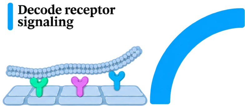
We build mimetic platforms that recreate key features of cellular interfaces to study how cells respond to chemical and mechanical cues during cell–cell communication. Our platforms span the nanoscale to the mesoscale, allowing us to answer a broad variety of biological questions. We use lithographic techniques to present ligands to cells at defined densities and in different spatial patterns to investigate how ligand density and arrangement influence receptor clustering, signaling, and cellular responses.
We use super-resolution microscopy to map receptor organization at the nanoscale and live-cell imaging to track how receptors cluster and reorganize over time. Optogenetic tools allow us to control when and where signaling occurs and to examine how individual cells respond. To study receptor mechanosensing, we vary the surface topography and mechanical properties of our platforms. By tailoring the ligands and scaffold properties, we can adapt these platforms to any receptor of interest.
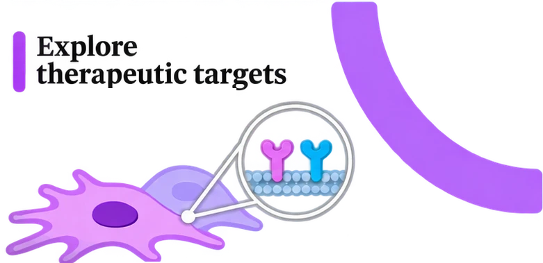
We study plexins and their semaphorin ligands as potential therapeutic targets in cancer. During nervous and cardiovascular system development, semaphorin signals help guide cell movement by activating plexin receptors. Through small GTPase signaling, receptor activation can influence cell shape, migration, and proliferation. In cancer, this guidance system has been linked to local invasion in glioblastoma and to metastatic spread in breast, pancreatic, and colorectal cancers.
The mechanisms by which plexins regulate cancer cell behavior remain poorly understood. One open question is how the same guidance cue can elicit different responses across cell types and tissue environments. We investigate how semaphorin presentation influences the organization of plexins at the cell membrane and how this organization affects cell motility. Our goal is to identify signaling interactions that could be targeted to limit invasion or metastatic spread in different cancer types.

We use direct laser writing via two-photon polymerization, a high-resolution 3D printing technique, to fabricate biodegradable scaffolds for local drug delivery in cancer. We investigate how scaffold composition and geometry influence contact with the surrounding tissue, scaffold degradation, and drug release kinetics. In collaboration with Prof. Philipp Backhaus at the European Institute for Molecular Imaging in Münster, we are studying the local delivery of STING agonists in a glioblastoma model. In the longer term, we aim to adapt these scaffolds to carry other therapeutic molecules and extend this work to nano- and microscale robots that can be guided to specific sites for controlled drug release in situ.
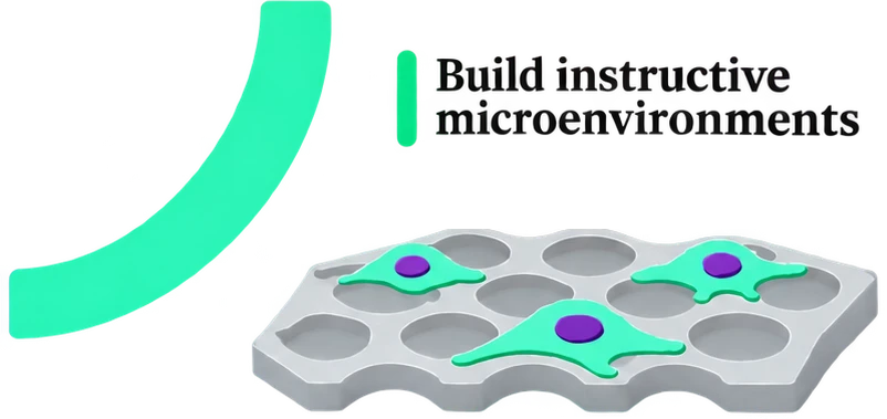
How does the physical environment of a tumor shape the way cancer cells respond to signals? In the tumor microenvironment, cells interpret chemical cues while interacting with neighboring cells and moving through three-dimensional tissue. We use 3D printing to build scaffolds that recreate selected features of this environment. These scaffolds allow us to study the interface between living and non-living matter by examining how cells attach, spread, and migrate in response to the geometry and surface properties of the surrounding material.
Our long-term vision
Our long-term vision is to combine our understanding of plexin signaling with bioengineering to develop biodegradable nanodevices that can navigate the three-dimensional tumor microenvironment and, for example, deliver peptides designed to block semaphorin-plexin interactions at the surface of cancer cells.
Project: Decoding Plexin signaling by chemo- and mechanosignals

Project: Targeting Plexin signaling and its role in immune modulation
Project: Dynamic and spatiotemporal control of Notch and Plexin signaling for assembling precise multicellular structures

Project: Programmable 3D-printed scaffolds for local drug release in glioblastoma
Scroll sideways for more →